
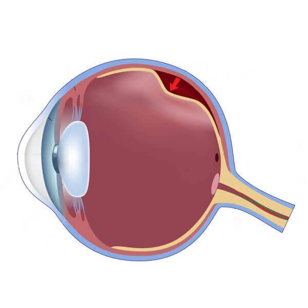
Retinal detachment is an emergency situation in which a thin layer of tissue at the back of the eye pulls away from its normal position. Retinal detachment separates the retinal cells from the layer of blood vessels that provides oxygen and nourishment. A break may be initially localized but without rapid treatment, the entire retina may detach, leading to vision loss and blindness.
If you use glasses or contact lenses to improve your sight, consider LASIK vision correction for the benefits.
There are several methods of treating a detached retina:
Vission Tip: Incase you have any floaters or flashes, please visit your doctor on a regular basis.

Common symptoms of retinal detachment include sudden flashes of light, floaters (small spots or threads in vision), blurred vision, reduced side vision, or a shadow/curtain appearing over part of the eye. Retinal detachment is considered a medical emergency and requires immediate treatment.
Retinal detachment can occur due to ageing, eye injuries, severe nearsightedness (myopia), diabetes, previous eye surgery, or retinal tears. In some cases, fluid builds up behind the retina and causes it to separate from its normal position.
Mild retinal tears may sometimes be treated with laser procedures, but most retinal detachment cases require surgery to prevent permanent vision loss. Treatment options may include retinal laser treatment, vitrectomy, or other retinal procedures recommended by a retina specialist.
Vision recovery depends on how quickly the condition is treated and the severity of the detachment. Early diagnosis and prompt retinal detachment surgery improve the chances of restoring vision and preventing permanent blindness.
Our Commitment to clinical excellence is lead by our senior doctor, Dr. Himanshu Mehta, a world renowned ophthalmic surgeon, who has performed over 25000 refractive surgeries
2026 All Rights Reserved Vission Eye Center | Design & Developed By Acetrot


Call

Consult

